
While many of us assume that our brains ramp down during sleep, this is not the case. In fact, your brain is very active during rapid-eye-movement (REM) sleep, which facilitates dreaming and memory processing.
It was recently discovered that histaminergic neurons of the LH and medial preoptic area (mPOA) are essential orchestrators of arousal and sleep. These neurons project to wake-promoting cholinergic and noradrenergic neurons, as well as GABA and glutamate neurons.
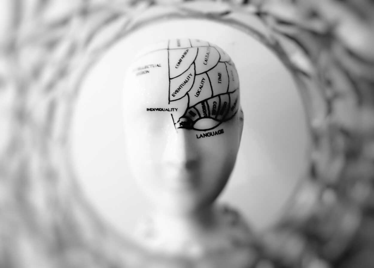
The Hypothalamus
The hypothalamus plays an important role in the coordination and regulation of many bodily systems including sleep and arousal, appetite, reproduction, and water intake. Abnormalities in these circuits can cause a variety of mental and physical issues – These data are the outcome of the service specialists’ inquiries https://sexgils.com.
Over the past several decades, lesion studies, pharmacological manipulations, and brain imaging have allowed researchers to identify and characterize the principal regions and neuronal populations involved in sleep/wake regulation. Traditionally, sleep/wake nuclei have been characterized as heterogeneous with intermingled neuronal populations that support different arousal states.
Despite this complexity, the lateral hypothalamus (LH) has emerged as an essential orchestrator of sleep and wakefulness. LH neurons express two alternatively spliced neuropeptides, hypocretin (orexin), that are released by cholinergic neurons in the basal forebrain and the brainstem. These neurons communicate with various wake-promoting populations in the brain including noradrenergic neurons of the locus coeruleus, dopaminergic neurons of the ventral tegmental area (VTA), and serotonergic neurons of the dorsal raphe nucleus and histaminergic neurons of the tuberomammillary nucleus.
The Pituitary
The pituitary gland is a pea-sized endocrine gland that is sometimes called the “master gland.” It secretes hormones that regulate the function of most other endocrine glands and control many major organs.
It also signals other glands and organs to produce hormones that affect important bodily functions, such as blood pressure, body temperature, hunger and thirst, urination, and heartbeat. The hypothalamus serves as the communications center for the pituitary gland by sending messages to it in the form of hormones. These hormones travel through a tube (the pituitary stalk) to the posterior pituitary gland.
The posterior pituitary gland consists of two distinct sections, the anterior and the posterior pituitary glands (also called the adenohypophysis and neurohypophysis). The anterior pituitary gland contains a large number of hormone-secreting epithelial cells. These cells secrete hormones including vasopressin and oxytocin, which are transported down the pituitary stalk to the posterior pituitary gland.
The Parabrachial Nucleus
Located in the dorsolateral pons near the lateral locus coeruleus and surrounded by the superior cerebellar peduncle, the parabrachial nucleus (PBN) has been shown to be essential for sensory processing. Genetically identified PBN neurons convey a variety of aversive stimuli including foot shock and taste memories to the central nucleus of the amygdala, the bed nucleus of the stria terminalis and other brain regions. Other PBN cells relay satiety, thirst or salt-appetite signals to the lateral hypothalamus and basal forebrain.
Over the past few years, it has become clear that the PBN has a large influence on sleep-wake dynamics. Using optogenetic and chemogenetic excitation/inhibition strategies, it has been shown that the lateral part of the PBN (LPBN) is the primary relay for arousal and sleep-promoting neurotransmitter signaling within the brainstem. The LPBN receives input from the hypothalamus, brainstem and thalamus through a dorsal pathway and then project to wake-promoting populations in the lateral hypothalamus and basal tegmental area (BTA). These projections include cholinergic neurons of the BTA and LC, noradrenergic neurons of the DA and DR and histaminergic neurons of the VTA.
The Locus Coeruleus
The locus coeruleus (LC) is a brain region involved in many neurobiological functions, including modulation of sleep-wake cycles, attentional control, responses to stress, memory formation and retrieval, and the production of the blue pigment neuromelanin, which gives this area its name. This area is also involved in lucid dreaming, which occurs when certain parts of the brain are stimulated during non-REM sleep.
The work of Moruzzi and Magoun showed that stimulation of LC neurons resulted in a brief cortical desynchronization that followed by high-voltage slow-wave activity associated with wakefulness. These results were supported by electrophysiological studies using sharpened tungsten electrodes that allowed for the recording of smaller neurons and their axons.
Later, immunohistochemical studies demonstrated that LC neurons send dense projections to the lateral hypothalamus, which releases the orexin peptide, and to serotonergic DRN circuitry, which controls arousal. This work was also supported by the finding that LC neurons are activated by sensory stimuli that are novel or unexpected, which is why they are considered to be part of the default mode and salience networks.
The Ventral Tegmental Area
While progress has been made in identifying neuronal populations that modulate sleep and wakefulness, determining how these diverse neurons coordinate their actions to stabilize arousal states remains a major challenge. It is now clear that fast gamma-Aminobutryic acid (GABA) and glutamate neurotransmission play important roles in sleep/wake regulation. In particular, VTA GABA neurons limit wakefulness via projections to arousal-promoting orexin neurons in the locus coeruleus and by inhibiting glutamatergic and DA neurons locally in the VTA.
The VTA is best known for harboring a group of cells referred to as DA neurons that are crucial in mediating reward consumption, learning, and memory, as well as addiction behaviors. However, it has only recently been realized that the caudal VTA contains a dense population of GABA neurons as well, which are also essential for regulating motivation and cognition. These GABA neurons are the focus of recent optogenetic studies.
The Preoptic Area
Nauta first used Fos imaging to identify a specific cell group in the rostral hypothalamus that was significantly active during sleep, and less so during wakefulness. He concluded that these neurons were responsible for sleep, and that lesion of the rostral hypothalamus produced sleeplessness.
Neurons in the ventrolateral preoptic nucleus (VLPO) show arousal-state dependent firing patterns and project to several other sleep/wake control regions, including the TMN and the basal forebrain. VLPO neurons secrete histamine, and their release is regulated by arousal state.
The VLPO contains subnuclei that differ in their cytoarchitecture, cellular signaling, and the types of neurons they contain. One, the intermediate nucleus, is found wedged between the lateral and medial preoptic nuclei and is distinguishable by variable expression of NPY- and CART-positive neurons. Another, the uncinate subnucleus, is distinguished by its cytoarchitecture and by its proximity to the medial preoptic nucleus. It is also characterized by its higher density of adenosine triphosphate (ATP)-generating neurons.


Leave a Comment